1Department of Forestry Technology, Federal College of Forestry, Jos, P.M.B. 2019, Bauchi, Plateau State, Nigeria, 2Montane Forest Research Station, Jos, Nigeria
ABSTRACT
Plants have been a source of medicine in pharmacopoeia. The African birch (Anogeissus leiocarpa) (DC) Guill and per. is an evergreen tree with traditional medicinal potentials against many ailments and diseases. Consequently, the phytochemical analysis and antibacterial activities of leaf extract of Anogeissus leiocarpa on Escherichia coli and Salmonella typhimurium were studied. Standard procedures were used to determine the phytochemical constituents of the extracts and susceptibility tests against the test organisms. The phytochemical analysis revealed the presence of active secondary metabolites such as alkaloids, tannins, phenols, steroids, saponins, flavonoids, and glycosides for plant extracts. A percentage yield of 48.29% was obtained for the leaf extract. The minimum inhibitory concentration (MIC) of the ethanolic extract of A. leiocarpa on E. coli and S. typhimurium was at 12.5 mg/ml and 6.25 mg/ml, respectively. The minimum bactericidal concentration was at 100 mg/ml for S. typhimurium and no effect was recorded for E. coli. The test organisms showed significant (P < 0.05) visible zones of inhibition at various concentrations of the extracts. The effectiveness of the different concentration on the tested organisms showed was comparable to the test drug, good alternative for the treatment of diarrhea fever in human.
Keywords: Anogeissus leiocarpa, Leaf extracts, Phytochemicals, Antibacterial
Plants form the basis of traditional medicine system which has been used since ancient time. Traditional medicine refers to health practices, approaches, knowledge and beliefs incorporating plant, animal and mineral-based medicines, spiritual therapies, manual techniques and exercises, applied singularly or in combination to treat, and prevent illnesses or maintain well-being.[1] The use of plant parts in the treatment of human diseases is as old as the disease itself and herbal medicines were the major form of medicine in Nigeria. About 80% of the world population depends on traditional medicine for their primary health-care needs today and their derivatives.[2] Plants have been a source of medicine in pharmacopoeia. Herbal medicine can be used as an alternative to some commercial drugs.[3] Medicinal plants provide inestimable projections for new drug discoveries because of them matchless availability of chemical range. The practice of herbal medicines in Asia signifies a long antiquity of human interactions with the environment.[4] Medical uses of plants range from the administration of the roots, barks, stems, leaves, and seeds to the use of extracts and decoction from the plants.[5] Medicinal plants are used as excellent antimicrobial agents because they possess a variety of chemical constituent in nature. Recently, much attention has directed toward the secondary metabolites and biologically active compounds from popular plant species.[6]
Anogeissus leiocarpa belongs to the family Combretaceae.[7] It is a deciduous tree that is sparsely distributed and sourced due to its trado-medical importance. It can grow to a height of up to 30 m, but typically 15–18 m with light green foliage and wider trunk at the base which sometimes striped. A. leiocarpa is commonly known as African Birch, Marke in Hausa, Atara in Igbo, and Orin – odan in Yoruba.[8] Conventionally, the root of the A. leiocarpa when used as chewing stick is known to have antibacterial effects on Lactobacillus spp.[9] The extracts of the plant in combination with that of Xanthoxylum gilletti mixed with Citrus juice revealed efficacy on HIV-associated opportunistic infections and complications.[10] A. leiocarpa demonstrated antimicrobial activity against a variety of viruses, malaria parasite, and some bacteria.[11] It is also used in traditional medicine as a remedy for many ailments, livestock sand man, which include helminthosis, schistosomiasis, leprosy, diarrhea, and psoriasis.[12]
Escherichia coli is a Gram-negative facultative anaerobic, rod shaped bacterium of genus Escherichia which can cause food poisoning in their host and cause various infection and become life treating. E. coli cause severe infection in human such as diarrhea, stomach cramping, and abdominal pain.[13] Salmonella typhimurium is a Gram-negative rod-shaped bacterium which belongs to the family Enterobacteriaceae which causes food poisoning in human, resulting in gastroenteritis in human causing serious infection such as diarrhea, fever disorder, and abdominal pain.[14] S. typhimurium has reported to have killed over 600,000 people annually all over the world. It is a deadly bacterial disease that causes typhoid fever, and it is transmitted through food and water.[15]
There is an increasing global concern on the emergence of antimicrobial resistant bacterial strains.[16] This has resulted to reduction in the effectiveness of current drugs and significantly causes treatment failure of infection. In many developing countries, the available drugs are costly and beyond the reach of common man.[1] High cost and accessibility to cheaper effective drugs against S. typhimurium and E. coli related ailments remain the major impediment to healthcare. The use of ethnobotanicals as antimicrobials stems from limitations due to toxicity, side effects, and multiple resistances of microorganisms associated with contemporary antimicrobial therapy.[17,18] The aforementioned challenges with orthodox option,[12,17] consequently stirs up the need to investigate the phytochemical screening and antibacterial activities of leaf extracts A. leiocarpa against E. coli and S. typhimurium.
The leaves of Anogeissus leiocarpa were collected from Tilden-Fulani, Toro LGA of Bauchi state. Following identification in the herbarium of the Federal College of Forestry, Jos, the materials were washed with distilled water and shade-dried for 2 weeks, to maintain its compositional integrity.[18] The dried materials were pulverized, using mortar and pestle, stored in air tight sterilized glass bottle until used.[19] One hundred and twenty grams of the pulverized substrate were dissolved in 80 ml of ethanol and allowed to stand for 24 h, thereafter filtered, using Whatman filter paper. The filtrate obtained was evaporated to dryness, using hot air oven at 37 (37°C) and stored in a refrigerator (4°C) until used.
The percentage yield extract was calculated using the formula below:
Where:
X1=Weight of empty beaker
X2=Weight of beaker + final dried extract
WSE = Weight of sample before extraction.[20]
The presence of some secondary metabolites in pulverized plant materials was determined using Standard methods.[21] This involves:-
One gram (1.0 g) of the leaf extract was dissolved in 10 ml of distilled water, shaken vigorously for 30 s and allowed to stand for 30 min. A honey comb-like froth formed for more than 30 min indicated the presence of saponins.
Two (2.0 ml) of acetic anhydride was added to 2 ml of the extract in a test tube. One milliliter of conc. H2SO4 was added down the side of the tube. A blue-green coloration indicated the presence of steroids.
Half gram (0.5 g) of the leaf extract was dissolved in 2.0 ml of chloroform, followed by addition of 3.0 ml of conc. H2SO4. A reddish brown coloration at the interface revealed the presence of terpenoids.
This involved sodium hydroxide test. Five drops of aqueous NaOH were added to 5 ml of each extract, a yellow coloration shows the presence of flavonoids.
Into 1.0 ml of the leaf extract in a conical flask was added 2.0 ml of Fec13. A dark green color gave a positive test for tannins.[22]
Two drops of the Dragendorffs reagent were added to 2.0 ml of the extract. A rose red precipitate indicates the presence of alkaloids, while Wagner’s Test with two drops of the Wagner’s reagent was added to 2.0 ml of the extract. A brown/reddish brown precipitate indicates the presence of alkaloids.
Into a test tube containing 0.5 g of the sample was added 2.0 ml of acetic anhydride. This was followed by addition of 2.0 ml H2SO4. A color changed from violet to blue-green indicated the presence of steroids.[23]
Culture of the test bacteria (E. coli and S. typhimurium) was obtained from National Veterinary Research Institute, VOM, Jos South LGA, Plateau State, Nigeria. Their identities were confirmed using cultural morphological and biochemical tests.[24] The bacteria isolates were maintained on nutrient agar at 4°C.
The antimicrobial effects of the ethanolic leaf extract of A. leiocarpa were determined using agar diffusion methods according to.[20] Sterilized nutrient agar was poured into Petri dish and allowed to set. Nutrient broth inoculated with the test bacteria was poured into the already set Petri dish and uniformly distributed. The control was introduced at the center, allowed to diffuse for 2 h at room temperature and incubated at 37°C for 24 h. The zone of inhibition (ZOI) due to the various responses of the test organisms at various concentrations of the extracts was measured with a transparent ruler. This measured the degree of sensitivity.
The MIC of the extracts against the test organisms was determined using agar diffusion method on the test organisms.[18,25] The test was performed at five concentrations of the leaf extract (100, 50, 25, 12.5, and 6.25 mg/ml), employing double dilution of extract infusion broth up to the fifth dilution. The test organisms were inoculated into each brain heart (or malt agar) and incubated overnight, after which 0.1 ml was added to all test tube and preparations were incubated at 37°C for 24 h. After incubation, a loop full from each tube was sub-cultured on nutrient agar to see if bacteria growth was inhibited. Growth of bacteria on solid media indicated that particular concentration of the extract was unable to inhibit the bacteria. The MIC was recorded as the lowest concentration of an antimicrobial agent that inhibited the visible growth of a microorganism after incubation for 24 h at 37°C.[26]
The MBC was determined by sub-culturing all the test tubes in each set that showed no turbidity or visible growth during MIC tests. The test tubes were incubated for 24 h at 37°C. The MBC was recorded as the lowest concentration that shows no growth or absence of growth after sub-culturing was considered as the MBC.[26]
Data obtained were subjected to analysis of variance, using SPSS version 16, to determine the level of significance, while significant means were separated using least significant difference.
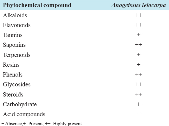
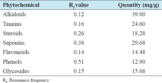
The percentage yield of the ethanolic extracts of the plant leaves showed percentage yields of 48.29% (Table 1). The phytochemical screening of ethanolic leaf extracts revealed the presence and variations in alkaloids, glycosides, steroids, anthraquinines, phenols, tannins, saponins, resins, terpenoids, flavonoids (Table 2), and carbohydrates based on qualitative assessment. However, the quantitative analysis based on resonance frequency revealed was in the order of alkaloids (39.00 mg/g) >saponins (29.68 mg/g) >tannins (24.60 mg/g) >steroids (18.28 mg/g) >glycosides (15.68) >flavonoids (14.48) >phenols (12.90 mg/g) (Table 3).
Table 1: Percentage yield of plant extracts
Table 2: Phytochemical constituents of leaf extracts of Anogeissus leiocarpa
Table 3: Quantitative phytochemical constituents of leaf extracts of Anogeissus leiocarpa
The MIC of leaf extract of A. leiocarpa against E. coli and S. typhimurium was 12.5 mg/ml and ≤6.25 mg/ml, respectively, after 24 h of incubation (Table 4). After a follow-up assay, the MBC was 100 mg/ml and >100 mg/ml for S. typhimurium and E. coli, respectively.
Table 4: Minimum inhibitory concentration and minimum bactericidal concentration of leaf extracts of Anogeissus leiocarpa against test organisms
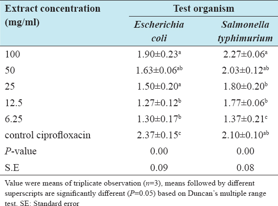
The ZOI of the leaf extract of A. leiocarpa on the test organisms showed that the test organisms were susceptible to the crude extracts at different degree and their susceptibility was concentration dependent. The ZOI varied with the concentrations (100 mg/ml, 50 mg/ml, 25 mg/ml, 12.5 mg/ml, and 6.25 mg/ml) of the ethanolic extracts of leaf of A. leiocarpa. The ZOI increased with increase in concentration of the extracts. S. typhimurium had higher values of ZOI with leaf extract than E. coli, while the contrary was the case with the stem bark (Table 5).
Table 5: Average zone of inhibition (cm) of the leaf extract concentrations of Anogeissus leiocarpa on the test organisms
The ethanolic leaf extract of A. leiocarpa revealed the presence of some phytochemicals that inhibited the growth of some microorganisms. This is consistent with the findings of Nweze et al.,[27] who found that various extracts of the leaf and stem bark of A. leiocarpa possessed active phytochemical constituents which inhibit the growth of isolates. This result also corroborated the findings of Edeoga et al.[28] and Zumbes et al.[29] The quantitative phytochemical analysis of leaf extract of Anogeissus leiocarpa showed the quantitative value and resonance frequency values of the active principles present in the plant extract. According to Ogundana et al.[30] and Nwinyi et al.,[31] the test plant contains phytochemical constituent, whose antioxidant could be the responsible for the antibacterial activities.[32]
The MIC ranged from 6.25 to 12.5 mg/ml was lower than the earlier reported values by Mann[33] and Timothy et al.[34] These were attributed to presence of the active phytochemical compounds.[35] The inhibition of the microbes by these secondary metabolites indicated their potentials in the treatment of diseases caused by the organisms. The ethanolic leaf extracts had bactericidal effect at 100 mg/ml for S. typhimurium and no effect was recorded for E. coli, indicating relative effects of concentrations, and consequently suggesting the higher concentrations than the selected range for E. coli. However, lethal effects on S. typhimurium are consistent with reported findings.[29]
The antibacterial activities of the ethanolic leaf extracts of A. leiocarpa and standard antibiotic on the test organisms showed effectiveness at varying levels and are concentration dependent. The observed diameter of the zones of inhibition indicated comparable effectiveness of the leaf extract with the control drug (ciprofloxacin) against the tested organisms especially S. typhimurium. This corroborated the report of Adejumobi et al.,[32] where the extracts inhibited the growth of microorganisms under both in vitro and in vivo conditions. The effects of concentration of ethanolic leaf extract of A. leiocarpa were significant (P < 0.05) on the ZOI against E. coli and Salmonella typhimurium. This is in line with the work of Adejumobi et al.,[36] showing that the ethanolic extract had varying activities against a wide range of pathogenic bacteria.
This study revealed the antibacterial potentials of leaf extract of A. leiocarpa on E. coli and S. typhimurium, with better efficacy against the later than the former. It is recommended that further studies be carried with higher concentrations of the leaf extract, while other vegetation parts of the test plant are considered.
The assistance of all the laboratory staff is highly appreciated.
There is no conflict of interests during this study.
1. World Health Organization. Traditional Medicine. Geneva:World Health Organization;2003. Available from:http//www.who/m/medical/centre/factsheet/ts/34/en/pirria/html. [Last accessed on 2020 Nov 22].
2. Mann A, Amupitan JO, Oyewale AO, Okogun JI, Ibrahim K. An ethnobotanical survey of indigenous flora for treating tuberculosis and other respiratory diseases in Niger State, Nigeria. J Phytomed Ther 2007;12:1-12.
3. Anyamene CO, Ezeadila JO. Antibacterial activity of water, ethanol and methanol extracts of Ocimum gratissimum, Vernonia amygdalina and Afromomum melegueta. J Appl Sci 2010;13:8940-8.
4. Sasidharan S, Chen Y, Saravanan D, Sundram KM, Latha LY. Extraction, isolation and characterization of bioactive compounds from plants'extracts. Afr J Tradit Complement Altern Med 2011;8:1-101.
5. Ogbulie JN, Ogueke CC, Nwanebu FC. Antibacterial properties of Uvaria chamae, Congronema latifolium, Garcinia kola, Vemonia amygdalina and Aframomium melegueta. Afr J Biotechnol 2007;6:1549-53.
6. Prince L, Prabakaran P. Antifungal activity of medicinal plants against plant pathogenic fungus Colletotrichum falcatum. Asian J Plant Sci Res 2011;1:84-7.
7. Hennenberg KJ, Geotze V, Minden DT, Porembsk S. Size-class distribution of Anogeissus leiocarpa (Combretaceae) along forest savannah ecotones in Northern Ivory coast. J Trop Ecol 2005;21:273-81.
8. Victor YA. In vitro assessment of antioxidant and antimicrobial activities of methanol extracts of six wound healing medicinal plants. J Nat Sci Res 2013;3:74-82.
9. Owoseni AA, Ogunnusi T. Antibacterial effect three selected chewing sticks extracts on Lactobacillus spp. Into J Trop Med 2006;3:103.
10. Kwame TA, Stephen KW, Anthony FK, Jeffery W. Finora A. Compositions Comprising Natural Agent for the Treatment of HIV-Associated Opportunities Infection and Complication and Method for Prepating and Using Composition Comprising Natural Agents;2005. Available from:http://www.freepalentsonline.comly2005/0266105. [Last accessed on 2018 Oct 12].
11. Taiwo O, Xu H, Lee S. Antibacterial activities of extracts from Nigerian chewing sticks. Phytother Res 1999;13:675-9.
12. Onyeyili PA. Anthelminthic Efficacy of Some Plants used in Ethnoveterinary Practices in the Arid Zone of North Eastern Nigeria RGA No. 28, Project Report;2000. 21.
13. Amin OM. Perspectives on gastro-intestinal pathogenic bacteria infections in humans. EC Microbiol 2019;15:1173-85.
14. Rosenberger CM, Scott MG, Gold MR, Hancock RE, Finley, RB. Salmonella typhimurium infection and lipopolysaccharide stimulation induce similar changes in macrophage gene expression. J Immunol 2000;164:5894-904.
15. Yasin N, Jabeen A, Nisa I, Tasleem U, Khan H, Shah FM, et al. A review:Typhoid fever. J Bacteriol Infect Dis 2018;2:1-7.
16. Hancock EW. Mechanisms of action of never antibiotic for gram-positive pathogens Lancet Infect Dis 2005;5:209-18.
17. Salih RR, Gibreel HH, Ahmed DM, Hammad ZM, Ahmed AI. Antimicrobial effect of aqueous and ethanolic leaves extracts of Ziziphus species against animal bacterial pathogens. Curr Trends For Res2019;3:1-6.
18. Chomini MS, Peter MK, Ameh M, Chomini AE, Bassey EA, Ayodele AO. Phytochemical Screening and Antibacterial Activities of Aframomum melegueta (K. Schum) Seed Extracts on Salmonella typhi and Klebsiella pneumoniae. JAppl Sci Environ Manage 2020;24:1419-24.
19. Ncube NS, Afolayan AJ, Okoh AI. Assessment techniques of antimicrobial properties of natural compounds of plant origin:Current methods and future trends. Afr J Biotechnol 2008;7:1797-806.
20. Ardzard SS, Yusuf AA, Muhammed M, David S, Odugba M, Okwon AE. Combine antibacterial effect of Moringa oleifera leaves extract and honey on some bacteria associated with wounds and gastroenteritis. J Adv Med Pharm Sci 2009;3:16-23.
21. Parekh J, Jadeja D, Chanda S. Efficacy of aqueous and methanol extracts of some medicinal plants for potential antibacterial activity. Turk J Biol 2005;29:203-10.
22. Ikeyi A, Ogbonna A, Eze F. Phytochemical analysis of paw-paw (Carica papaya) leaves. Int J Life Sci Biotechnol Pharm Res 2013;2:347-51.
23. Ghamba PE, Balla H, Goje LJ, Halidu A, Dauda MD. In vitro antimicrobial activities of Vernonia amygdalina on selected clinical isolates. Int J Curr Microbiol Appl Sci 2014;3:1103-13.
24. Akimnibosun HA, Akinnibosun FI, Adieme BC. Bio therapeutic potential of aqueous an ethanol extracts of leaves on some vegetable gram negative bacteria. Bot Environ Sci JTrop2009;6:3337.
25. Cheesbrough M. Antibacterial Sensitive Affecting. District Laboratory Practice in Tropical Counties, Part 2. Cape Town, South Africa:Cambridge University Press;2000. 132-43.
26. Cheruiyot KR, Olila D, Kateregga J. In vitro antibacterial activity of selected medicinal plants from Longisa region of Bomet district. Kenya Afr Health Sci 2009;9:542-6.
27. Nweze EI, Okafor JI, Njoku O. Antimicrobial activities of methanolic extracts of Trema guineensis (Schumm and Thorn) and Morinda lucida Benth used in Nigerian herbal medicinal practice. J Biol Res Biotechnol 2004;2:39-46.
28. Edeoga HO, Okwu DE, Mbaebie BO. Phytochemical constituents of some Nigerian medicinal plants. Afr J Biotechnol 2005;4:685-8.
29. Zumbes HJ, Belenu TO, Onwuliri FC. In vitro antibacterial activity of Anogeissus leiocarpa leaf extracts on some bacteria associated with diarrhea. Int J Nat Appl Sci 2007;3:53-6.
30. Ogundana OT, Gunlaria O, Farombi OE. Morindalucida;antionidant and reducing activities of crude methanolic stem bark extracts. Adv Nat Appl Sci 2008;2:49-54.
31. Nwinyi OC, Chinedu NS, Ajani OO. Evaluation of antibacterial activities of Pisidum guajava and Gongronema latifolium. J Med Plant Res 2008;2:189-92.
32. Adejumobi JA, Ogundiya MO, Kolapo AL, Okunada MB. Phytochemical composition and in vitro antimicrobial activity of Anogeissus leiocarpa on some common oval pathogen. J Med Plants Res 2008;2:193-6.
33. Mann A. Evaluation of antimicrobial activity of Anogeissus leiocarpus and Terminalia auicenniodes against infection diseases prevelent in Hospital environment in Nigeria. J Microbiol Res 2012;2:6-1.
34. Timothy SY, Mashi FI, Helga BI, Galadima IH, Midala TA. Phytochemical screening, antibacterial evaluation and in vitro spasmodic effect of the aqueous and ethanol leaf and stem bark extracts of Anogeissus leiocarpus (DC) Guill and Perr. Asian J Pharm Sci Technol 2015;5:302-8.
35. Umar HY, Muhammad AI. Antibacterial activities of leaves extract of Anogeissus leiocarpa and Vitex doniana against some bacteria. Am J Innov Res Appl Sci 2015;1:384-8.
36. Bereksi MS, Hassaïne H, Bekhechi C, Abdelouahid DE. Evaluation of antibacterial activity of some medicinal plants extracts commonly used in Algerian traditional medicine against some pathogenic bacteria. Pharmacogn J 2018;10:507-12.