large

Phytochemical screening and antimicrobial effects of leaf extracts of Acanthospermum hispidum D.C. on four Salmonella strains

Stephen Meyiwa Chomini*, Theopthilus Aimuamwosa Erhabor, Chidiebere James Ukanyirioha, Joy Abiola Imoh, Yahaya Sadiku, Martins Ukwadi, Stephen Adebayo Popoola, Nanle Janfa, Mundi Junior Francis, Collina Kambai, Augustina Aigbe Bello

Department of Forestry Technology, Federal College of Forestry, Jos, Plateau State, Nigeria

Address for correspondence: Stephen Meyiwa Chomini, Department of Forestry Technology, Federal College of Forestry, Jos, Plateau State, Nigeria. E-mail: stevemchoms@gmail.com
Submitted: 29-04-2021, Accepted: 03-05-2021, Published: 30-06-2021

ABSTRACT

Herbal medicines are being used as an alternative to some orthodox drugs for the treatment of a wide range of diseases and their efficacies have been mainly attributed to inherent secondary metabolites. This study, therefore, aimed at investigating the phytochemical constituents and antibacterial effects of methanolic leaf extract of Acanthospermum hispidum on four Salmonella species. Standard methods were used to assay the phytochemical constituents. Five concentrations: 62.5 mg/ml, 125 mg/ml, 250 mg/ml, 500 mg/ml of the extracts, and 0.1 ml of 1.7 mg/ml ciprofloxacin (as control drug) were screened for antibacterial activities against Salmonella typhi, Salmonella typhimurium, Salmonella Gallinarum, and Salmonella paratyphi, using agar well diffusion methods. The leaf extract showed the presence of alkaloids, anthraquinones, resins, tannins, flavonoids, carbohydrate, phenols and protein, and amino acids, while saponins were absent, with a percentage yield of 48.29%. The average inhibition zone against the test organisms increased with extract concentrations, with S. paratyphi and S. typhi having highest (16.0 mm) and lowest (9.0 mm) values under 500.0 mg/ml. The control drug had significant effects over leaf test concentrations. All test strains had 125 mg/ml and 250 mg/m as minimum inhibitory concentration (MIC) and minimum bactericidal concentration (MBC), except S. typhi with 250 mg/ml and 500 mg/ml, respectively. The antibiotic power (MBC/MIC ratio) of the test extract was 2.0, indicating a bactericidal effect, while the antibiotic potential ranged from moderate to strong. These findings elucidated the potential of the leaf as possible good alternative for the treatment of gastrointestinal infection in human.

Keywords: Acanthospermum hispidum, bactericidal, inhibitory, Salmonella


INTRODUCTION

Medicinal plants are the richest bioresources of traditional medicine, food supplements, folk medicines, and pharmaceutical raw materials for synthetic drugs.[1] Herbaceous plants have been employed for the treatment of a wide range of diseases, with recorded efficacies mainly attributed to inherent secondary metabolites they possess.[2] Many of these natural compounds such as flavonoids, alkaloids, terpenoids, carotenoids, and lignin are biodegradable, environmentally friendly with low record of toxicity. Salmonellosis, enteritidis, and other gastrointestinal illnesses remain a global public health concerns.[3] Many antibiotics are becoming less effective, due to increasing resistance by pathogenic microbes.[4,5] Phytomedicine disease control is increasing in developing countries.[6,7]

Leaf extracts of different plant species have been reported to show potential antimicrobial effects. Azadirachta indica, Mimosa pudica, Chromolaena odorata, and Acacia nilotica have indicated growth inhibitory effects against Bacillus subtilis, Staphylococcus spp., and Streptococcus spp.[8,9] Vernonia polyanthes extract gave a positive inhibitory effects against Leishmania strains while alkaloid extract of Phyllanthus discoideus was reported to show strong inhibition against pathogenic bacteria such as Escherichia coli, Enterococcus faecium, Pseudomonas aeruginosa, Staphylococcus aureus, and Mycobacterium smegmatis.[10] Methanolic leaf extracts of Mentha piperita (peppermint), Cymbopogon citratus (lemongrass), Allium sativum (garlic), Syzygium aromaticum (clove), and Zingiber officinale (ginger) have established satisfactory antimicrobial tendencies against S. aureus.[10]

Acanthospermum hispidum D.C. bristly starbur, goat’s head, kashin-yaawoo (Hausa) and yaawoo (“Ron” tribe),[11] belongs to the family Asteraceae. It is an annual plant with hairy stem. The leaves are elliptic, obovate and 1.5 cm–7.0 cm long, although a length of up to 11.5 cm long has been reported. It bears yellow flowers. The fruits are flattened, triangularly shaped and spiny and 5 cm–10 cm in length. These fruits are covered with stiff, hooked hairs and have either a straight or curved pair of spines at the top.[12]

A number of attempts have been reported on the use of A. hispidum as an alternative option against infectious diseases. According to Chakraborty et al.,[12] a 50% aqueous ethanolic extracts of A. hispidum possess antitumor and cancer potentials. Okoro et al.[13] reported significant antimicrobial activities of acetone extract of A. hispidum against some clinical isolates, with inhibition zones ranging from 21 to 31 mm and minimum inhibitory concentration (MIC) between 1.25 and 20.00 mg/ml. Traditional applications for cases of jaundice, malaria, fever, stomach ache, constipation, and viral infections have been documented.[14,15]

This paper focuses on an attempt to assess the phytochemical and antimicrobial activity of methanolic leaf extracts of A. hispidum D.C. on four strains of Salmonella.

MATERIALS AND METHODS

Collection and Methanolic Extraction of Test Plant Materials

Collected leaves of A hispidum were identified in the Herbarium of the Federal College of Forestry, Jos. They were rinsed with distilled water and air-dried for 3 weeks under shade, thereafter pulverized using mortar and pestle, and kept aseptically in an airtight cellophane bag until use.[16]

Thirty grams of pulverized sample were macerated in 300 ml of 70% methanol for 48 h. After filtering with Whatman No. 1 filter paper, the filtrates were evaporated to dryness using hot air oven at 70°C. The extracts in airtight bottles were stored in desiccators until used.[16,17]

The percentage yield of the extract was calculated using the formula below as described by Ardzard et al.[17]

thumblarge

Where, x1= weight of empty beaker; x2= weight of empty beaker + final dried extract.

Phytochemical Screening and Stock Test Concentration from the Plant Extracts

The presence of some basic secondary metabolites in the pulverized plant materials was determined using standard methods.[18-20] 3.0 g of the extracts were dissolved in 6.0 ml of dimethyl sulfoxide.[17,21]

Antimicrobial Studies

Source and standardization of test organisms

Previously characterized clinical isolates of Salmonella typhi, Salmonella typhimurium, Salmonella paratyphi, and Salmonella gallinarum were obtained from National Veterinary Research Institute, Vom, Plateau state, Nigeria. Routine protocols of sterilization were observed for all glass wares wire loop and cork borers. While work bench surfaces were disinfected using Dettol.[16,17] The inoculums of the test organisms were standardized using the McFarland Nephelometer methods described by Albert et al.[22] The turbid solutions obtained by this protocol were kept on the work bench for use. The procedure ensured that bacterial suspension corresponds to 1.5 × 10⁵/mm bacterial suspension.

Culture Media Preparation

Nutrient agar (NA) was prepared according to manufactures instruction (Oxoid CM003, 28 in 1 L of distilled water). Twenty-eight grams of NA were weighed into a conical flask; 1000 ml of distilled water was added and capped. The medium was shaken to dissolution and sterilized at 121°C for 15 min.[17]

Determination of Antibacterial Activity

The antibacterial effects of methanol leaf extracts of A. hispidum were assessed using agar well diffusion methods as described by Perez et al.[23] and Olukoya et al.[24] Prepared NA plates were separately swabbed with the broth cultures of specific test organisms, allowed to diffuse at room temperature for 2 h, and incubated at 37°C for 24 h. Five equidistant wells (6.0 mm in diameter) were made in each plate, using a sterile cork borer. 0.1 ml NA was added to seal the bottom, while 62.5 mg/ml, 125 mg/ml, 250 mg/ml, and 500 mg/ml were, respectively, introduced into each well, using sterile Pasteur pipette. A 0.1 ml of 1.7 mg/ml ciprofloxacin as control was introduced into the well at the center. After 2 h of setting, the plates (in triplicates per organism) were incubated at 37°C for 24 h.

Diameters of the zones of inhibition were measured using a transparent ruler. The antibacterial activity of the extracts was expressed as mean diameter of zone of inhibition (mm) measured for each of the test organisms.

Determination of MIC

The MIC was carried out, using broth dilution method by Cowan and Steel.[25] 1.0 ml bacteriological peptone was poured into five test tubes (1–5). Into a test tube was 1.0 ml of the stock (extract) concentration added using Pasteur pipette. The content was thoroughly mixed to achieve even dilution and distribution. 1.0 ml of the mixture was withdrawn and transferred into tube 2 and from tube 2 into 3 and evenly mixed. This was repeated for all the tubes. Finally, 1.0 ml of the mixture was discarded from the last tube in the set. This process was done for all sets of the tubes. 0.1 ml inoculum of the test organism was inoculated into each tube, using sterile pipette, thoroughly mixed and incubated at 37°C for 24 h, after which they were examined for visible turbidity. Tubes which showed turbidity indicated microbial growth. The MIC, therefore, was represented the lowest concentration that prevented visible growth.[26]

Determination of Minimum Bactericidal Concentration (MBC)

The MBC was assessed by subculturing samples from all the tubes which tested negative for visible turbidity after MIC test. This was done by subculturing a loop full of contents from tubes with no growth, streaked over the surface of NA plates. The plates were incubated at 37°C for 24 h. Plates with no growth indicated bactericidal effect while the MBC was the lowest concentration at which no growth was observed after subculturing.[26]

Antimicrobial Indices

The effect of an antibiotic substance on a microorganism is measured by a number of indices relative to its MIC and MBC. The ratio of MBC/MIC describes the antibiotic power (AP).[27] According to Noumedem et al.,[28] if the ratio MBC/MIC is ≤4, the effect considered bactericidal, but the ratio is >4, the effect is described as bacteriostatic.

RESULTS AND DISCUSSION

The Phytochemical Constituents of A. hispidum

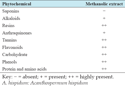
The phytochemical assay of methanolic leaf extracts of A. hispidum indicated the presence of alkaloids and anthraquinone, higher contents of resins, tannins, flavonoids, carbohydrate, phenol and protein, and amino acids, while saponin was absent [Table 1].

Table 1: Phytochemical component of methanolic extract of Acanthospermum hispidum D.C

thumblarge

Sensitivity Tests

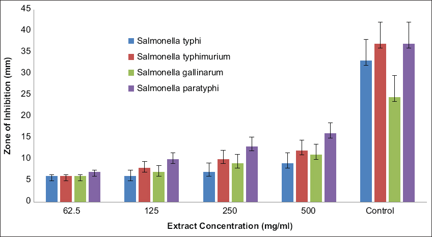
The sensitivity of the test microbial isolates to the leaf extract varied with microbial strains as well as the concentration of extract. There was a general increase in average inhibition zones (AIZ) with extract concentrations. S. typhi, S. typhimurium, S. gallinarum, and S. paratyphi gave 6.0–9.0 mm, 6.0– 12.0 mm, 6.0– 11.0 mm, and 7.0–16.0 mm, respectively, as the ranges of AIZ. S. paratyphi and S. typhi had the highest AIZ of 16.0 mm and 9.0 mm under 500 mg/ml [Figure 1]. The control drug ciprofloxacin (1.7 mg/ml) gave 33.0 mm, 37.0 mm, 24.5 mm, and 37.0 mm as the AIZ for S. typhi, S. typhimurium, S. gallinarum, and S. paratyphi, respectively. The analysis of variance indicated a significant difference in the effects of the test drug over the extracts of the test plant.

thumblarge

Figure 1: Average zones of inhibition (mm) for different concentrations of methanolic extract of Acanthospermum hispidum D.C. on the test organisms

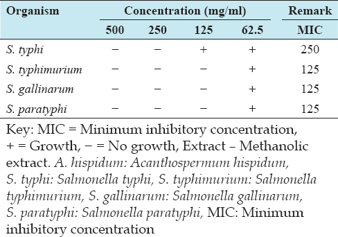
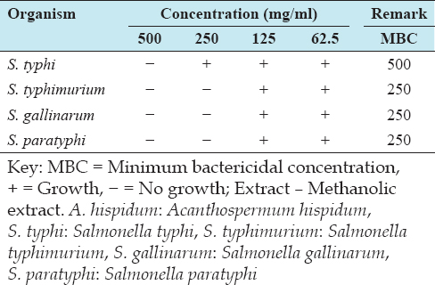
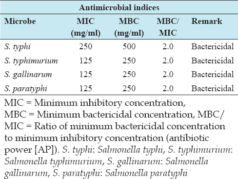
The methanolic leaf extract of A. hispidum recorded 125.0 mg/ml as MIC, against S, typhimurium, S. gallinarum, and S. paratyphi, while S. typhi had an MIC value of 250.0 mg/ml [Table 2]. The leaf extract of A. hispidum gave a MBC value of 250 mg/ml against S. typhimurium, S. gallinarum, and S. paratyphi. However, S. typhi recorded an MBC value of 500.0 mg/ml [Table 3].

Table 2: MIC of methanolic extract of A. hispidum D.C. on the test organisms

thumblarge

Table 3: MBC of methanolic extract of A. hispidum D. C. on the test organisms

thumblarge

Antimicrobial Parameters and Indices

The AP (MBC/MIC ratio) of the methanolic leaf extract evaluated for all the bacterial strains gave an AP value of 2.0, which is considered as bactericidal against the microbial isolates [Table 4].

Table 4: Antimicrobial parameters and indices

thumblarge

DISCUSSION

The relative presence of phytochemicals otherwise known as secondary metabolites in plants was reported to depend on the solvent types and the polarity of the extractants.[29] The phytochemical profile of A. hispidum obtained was similar,[13] except for the absence of saponins. They reported the presence carbohydrate, cardiac glycoside, saponins, alkaloids, anthraquinone, steroids, triterpene, tannins, and flavonoids and attributed the absence of saponins in acetone extract to its high hydrophilic affinity, readily dissolving in polar solvents. Usman et al.[30] indicated that tannins and flavonoids are established microbial growth inhibitors. Mishra et al.[31] have reported the propensity of flavonoids for microbial cell disturbance, inhibition of nucleic acid synthesis, and cell wall synthesis. Tannins were reported to form protein complex, which is lethal to bacteria, as it alters their biochemical process.[32] According to Maria et al.,[33] phenols exhibit antioxidant and enzyme inhibitory tendencies, while alkaloids have been found to cause structural and genetic imbalance as well as bacterial DNA cell wall damage.[34]

Sensitivity Tests

The antimicrobial effects of plant extracts are assessed on the basis of sensitivity response of test microorganisms. The tests were based on inhibition zones, bactericidal and bacteriostatic parameters. Firempong et al.[35] described the categorization of the zones of inhibition into four different benchmark ranges, namely, <5.0 mm, 5.0–10.0 mm, 10.0–20.0 mm, and ≥20.0 mm, as weak, moderate, strong, and very strong antibacterial potential, respectively. In this regard, the antimicrobial potentials of the extract varied with the microbial strains and concentrations. All extract concentrations had moderate antimicrobial potentials (MAP) on S. typhi. 62.5 mg/ml and 125.0 mg/ml gave MAP, while 250.0 mg/ml and 500.0 mg recorded strong antimicrobial potentials (SAP) against S. typhimurium. There was MAP of ≤250.0 mg/ml and SAP of 500 mg/ml against S. gallinarum. However, all extract concentrations showed SAP against S. paratyphi, except 62.5 mg/ml with MAP. This outcome was similar to the findings of Chomini et al.,[36] indicating antibacterial potentials of methanolic seed extracts of Aframomum melegueta on Klebsiella pneumoniae and S. typhi. The control drug showed very strong antibacterial potentials for on all the test microbes [Figure 1].

According to Nas et al.[37] and Ogodo et al.,[38] the low inhibition concentration value suggested better antibacterial activity, which reflected strong effects against the test organisms. Polar organic solvents have been reported to show better solubility than aqueous and consequently, higher bioactive constituents and better suppressive and lethal effects.[37] The ratio of MBC: MIC obtained for all test microbial isolates was 2.0, which according to Noumedem et al.,[28] was suggestive of bactericidal effects of the methanolic leaf extract of A. hispidum, since the ratio was ≤4.0.

CONCLUSION

The study revealed that methanolic leaf extracts of Acanthospermum hispidum (MLEAH) exhibited antibacterial activities against S. typhi, S. typhimurium, S. gallinarum, and S. paratyphi. The phytochemical screening of MLEAH showed the presence of alkaloids, anthraquinone, higher contents of resins, tannins, flavonoids, carbohydrate, phenol and protein, and amino acids, and the absence of saponins. The sensitivity tests indicated relative significant effect of the control drug over the test concentrations of MLEAH, with the MBC/MIC ratio showing a bactericidal effect across the tests bacterial strains as well as moderate-to-strong antibacterial properties on the test microbes. This elucidates its antimicrobial potentials for pharmaceutical industries and afforestation needs.

REFERENCES

1.  Ncube NS, Afolayan AJ, Okoh AI. Assessment techniques of antimicrobial properties of natural compounds of plant origin:Current methods and future trends. Afr J Biotechnol 2008;7:1797-806.

2.  Prakash D, Gupta KR. The antioxidant phytochemicals of nutraceutical importance. Open Nutr J 2009;2:20-35.

3.  Li XZ, Ma D, Livermore DM, Nikaido H. Role of efflux pump(s) in intrinsic resistance of Pseudomonas aeruginosa:Active efflux as a contributing factor to beta-lactamase resistance. Antimicrob Agents Chem 1994;38:1742-52.

4.  Levy SB, Marshall B. Antibacterial resistance worldwide:Causes, challenges and responses. Nat Med 2004;10:S122-9.

5.  Pamplona-Zomenhan BC, Pamplona CB, da Silva MC, Mimica LM. Evaluation of the in vitro antimicrobial activity of an ethanol extract of Brazilian classified propolis on strains of Staphylococcus aureus. Braz J Microbiol 2011;42:1259-64.

6.  Keita SM, Vincent C, Schmit JP, Belanger A. Essential oil composition of Ocimum basilicum, Ocimum gratissimum and Ocimum suave in the republic of guinea. Flavour Fragrance J 2000;15:339-41.

7.  Mann A, Salawn FB, Abdulrauf I. Antibacterial activity of Bombax buonopozense P. Beauv. (Bombacaceae) edible floral extracts. Eur J Sci Res 2011;48:627-30.

8.  Policegoudra RS, Abiraj K, Gowda DC, Aradhya SM. Isolation and characterization of the antioxidant and antibacterial compound from the mango ginger (Curcuma amada Roxb.) rhizome. J Chromatogr B Analyt Technol Biomed Life Sci 2007;852:40-8.

9.  Chattopadhyay D, Arunachalam G, Mandal AB, Sur TK, Mandal SC, Bhattacharya SK. Antimicrobial and anti-inflammatory activity of folklore:Mallotus peltatus leaf extract. J Ethnopharmacol 2002;82:229-37.

10.  Tyagi R, Sharma G, Jasuja ND, Menghani E. Indian medicinal plants as an effective antimicrobial agent. J Crit Rev 2016;3:69-71.

11.  Akobundu IO, Agyakwa CW. A Handbook of West African Weeds. 2nd ed. Ibadan, Nigeria:ITA;1998. 160-1, 194-5.

12.  Chakraborty AK, Gaikwad AV, Singh KB. Phytopharmacological review on Acanthospermum hispidum. J Appl Pharm Sci 2012;2:144-8.

13.  Okoro IO, Inegbedion AO, Okoro EO. Phytochemical screening and antibacterial activity of different solvent extracts of Acanthospermum hispidum Dc. Aerial parts. Niger Ann Nat Sci 2017;16:43-7.

14.  Mshana NR, Abbiw DK, Addae-Mensah I, Adjanouhoun E, Ahji RA, Ekpere JA, et al. Floristic Studies in Ghana. Ethiopia:OAU/STRC;2000. 101-2.

15.  Sumnerfield A, Keil GM, Mettenleiter TC, Rziha H, Saalmuller A. Antiviral activity of an extract from the leaves of the tropical plant Acanthospermum hispidum. Antiviral Res 2007;36:55-62.

16.  Apak L, Olila D. The in vitro antibacterial activity of Annona senegalensis Pers. and other plants-Ugandan medicinal plants. Afr Health Sci 2006;6:31-5.

17.  Ardzard SS, Yusuf AA, Muhammad M, David S, Odugba M, Okwon AE. Combine antibacterial effect of Moringa oleifera leave extract and honey on some Bacteria associated with wounds and gastroenteritis. J Adv Med Pharm Sci 2009;3:16-23.

18.  Sofowora A. Medicinal Plants and Traditional Medicine in Africa. 3rd ed. Nigeria:Spectrum Books Limited Ibadan;1993. 199-204.

19.  Evans WC. Trease and Evans Pharmacognosy. 15th ed. Netherlands:Elsevier Indian;2002. 137-93.

20.  Prashant T, Bimlesh K, Mandeep K, Gurpreet K, Harleen K. Phytochemical screening and extraction. A review. Int Pharm Sci 2011;1:98-108.

21.  Fleischer TC, Ameade EP, Sawer IK. Antimicrobial activity of the leaves and flowering tops of Acanthospermum hispidum. Fitoterapia 2003;74:130-2.

22.  Albert B, William JH, Keneth LH, Henry DI, Jean HS. Manual of Clinical Microbiology. 5th ed. Michigan:American Society for Microbiology, The University of Michigan;1991. 10.

23.  Perez C, Paul M, Bazerque P. Antibiotic assay by agar-well diffusion method. Acta Biol Med Exp 1990;15:113-5.

24.  Olukoya DK, Idika N, Odugbemi T. Antibacterial activity of extracts from some medicinal plants in Nigeria. J Ethnopharmacol 1993;39:69-72.

25.  Cowan ST, Steel KJ. Antibiotic Sensitivity in Cowan and Steel's Manual for Identification. London, New York:Cambridge University Press;1985. 24.

26.  Cheesbrough M. Antibacterial sensitivity testing. In:District Laboratory Practice in Tropical Countries. Part 2. Cambridge:Cambridge University Press;2000. 132-43.

27.  Yehouenou BB. Composition Chimique et Activites Antimicrobiennes D'extraits Vegetaux du Benin Contre les Microorganiques Pathogenes et Alterants des Denrees Alimentaires. These Unique de Doctorat. Benin:Universite d'Abomey-Calavi;2012. 221.

28.  Noumedem J, Mihasan M, Lacmata S, Stefan M, Kuiate J. Kuete V. Antibacterial activities of the methanol extracts of ten Cameroonian vegetables against Gram negative multidrug-resistant Bacteria. BMC Complement Altern Med 2013;13:26.

29.  Salama HM, Marraiki N. Antimicrobial activity and phytochemical analyses of Polygonum aviculare L. (Polygonaceae), naturally growing in Egypt. Saudi J Biol Sci 2010;17:57-63.

30.  Usman H, Abdulrahman FI, Ladan AA. Phytochemical and antimicrobial evaluation of Tribulus terrestris L. (Zygophylaceae) growing in Nigeria. Res J Biol Sci 2007;2:244-8.

31.  Mishra NN, Yang S, Sawa A, Rubio A, Nast CC, Yeaman MR, et al. Analysis of cell membrane characteristics of in vitro-selected daptomycin-resistant strains of methicillin-resistant Staphylococcus aureus. Antimicro Agents Chem 2009;53:2312-8.

32.  Shaimaa OH. Antibacterial activities of Iraqi propolis and its active components ex tracts on some bacterial isolates (in vitro study). World J Pharm Pharm Sci 2014;3:858-75.

33.  Maria BC, Vidya P, Manjula S, Maji J. Antimicrobial properties of coconut husk aqueous extraction cariogenic Bacteria. Arch Med Health Sci 2013;2:126-30.

34.  Firempong CK, Andoh LA, Akanwariwiak WG, Addo-Fordjour P, Adjakofi P. Phytochemical screening and antifungal activities of crude ethanol extracts of red-flowered silk cotton tree (Bombax buonopozense) and Calabash nutmeg (Monodora myristica) on Candida albicans. JMicrobiol Antimicrob 2016;8:22-7.

35.  Sarjono PR, Putri LD, Budiarti CE, Mulyani NS, Ngadiwiyana I, Kusrini D, et al. Antioxidant and Antibacterial Activities of Secondary Metabolite Endophytic Bacteria from Papaya Leaf (Carica papaya L.). London, United Kingdom:13th Joint Conference on Chemistry (13th JCC) IOP Conference Series:Materials Science and Engineering;2019. 012112.

36.  Chomini MS, Peter MK, Ameh M, Chomini AE, Bassey EA, Ayodele AO. Phytochemical screening and antibacterial activities of Aframomum melegueta (K. Schum) seed extracts on Salmonella typhi and Klebsiella pneumoniae. JAppl Sci Environ Manage 2020;24:1419-24.

37.  Nas S, Zage AU, Ali M. Phytochemical screening and antibacterial activity of seed extracts of Garcinia kola against pathogenic methicillin resistant Staphylococcus aureus. Bayero J Pure Appl Sci 2017;10:198-203.

38.  Ogodo AC, Nwaneri CB, Agwaranze DI, Inetianbor JE, Okoronkwo CU. Antimycotic and antibacterial activity of Aframomum melegueta seed extracts against Bacteria and fungi species from food sources. Cent Afr J Public Health 2017;3:44-50.