large

TNF-α regulated inflammatory pathway by Isopulegol in human lung adenocarcinoma (A549) cells through ROS generation

Periyannan Velu*

Scigen Research and Innovation Pvt. Ltd., Periyar Technology Business Incubator, Thanjavur, Tamil Nadu, India

Address for correspondence: Dr. Periyannan Velu, Scigen Research and Innovation Pvt. Ltd., Periyar Technology Business Incubator, Thanjavur, Tamil Nadu, India. Email: biovetrivel6@gmail.com
Submitted: 22-09-2020, Accepted: 06-11-2020, Published: 30-12-2020

ABSTRACT

In the present study, we have investigated the anti-adenocarcinoma potential of isopulegol against human lung adenocarcinoma (A549) cells. The cytotoxic effects of isopulegol on A549 were evaluated through MTT assay drug modified morphological analysis. The status of reactive oxygen species (ROS) generation, mitochondrial membrane potential (Δψm), and apoptosis induction in A549 cells were identified through DCFH-DA, Rh-123, and DAPI staining, respectively. The effect of isopulegol on the activities of antioxidant and lipid peroxidation (LPO) enzymes and expression patterns of apoptosis and inflammatory proteins were investigated using the RT-PCR and Western blotting technique in the lung cancer cells. The results confirmed that isopulegol has strong cytotoxicity against A549 cells. Moreover, increased ROS generation through induced apoptosis in A549 cells was confirmed through DCFH-DA, Rh-123, DAPI, and PI staining. Isopulegol drastically suppressed cell proliferation. Decreased levels of the antioxidant and increased level of LPO enzymes were also observed to reduce the level of inflammatory markers such as TNF-α and NF-kB. The Cox-2 expression was noted in isopulegol treated A549 cells. Inclusively, the isopulegol noticeably stimulated the cytotoxicity through increased ROS and apoptotic protein expressions in lung cancer A549 cells, thus suggesting its potency as a viable anticancer agent against human lung epithelial carcinoma (HLEC).

Keywords: A549 cells, apoptosis, inflammatory markers, isopulegol, lung cancer

INTRODUCTION

Lung adenocarcinoma is the most common subtype of non-small lung carcinomas, which has the highest mortality rate (30% or 0.1 million) globally.[1] At present, lung cancer is treated using several approaches, such as surgical elimination of tumors, chemotherapy, radiotherapy, and therapeutic medications, but in some cases, such approaches may fail to improve the condition of sarcoma patients, leading to death.[2] Even though pulmonary sarcoma is currently largely treated by surgical exclusion and medications, the success rate is low, with failure to improve the health condition of suffering patient, reported. Moreover, medical data showing that <15% of lung cancer patients benefited of the aforementioned treatments by surviving for ≥5 years.[3,4]

The chemotherapy is an extensively utilized treatment strategy of sarcoma in diminishing the number of malignant cells through cytotoxic actions of chemotherapeutic agents. Typically consecutive chemotherapeutic phases are used to reduce the recurrence of malignancy and to enhance treatment success. Conversely, in some cases, the chemotherapeutic strategy fails to reduce the malignant cells usually at the initial stage of treatment, due to drug resistance that may have been attained by the cancer patients. In addition, the existing chemotherapeutic strategy has less accuracy and results in significant toxicity to the normal cells, leading to a low survival rate of patients in many cases. Consequently, the ineffectiveness of existing chemotherapeutic strategy has urged the requisites of the development of novel and effective treatment approaches.[5-7]

Apoptosis is an essential process for the sustained development of living things; simply, it is the regeneration of cells and depends on the intracellular system.[8] Usually, two processes contribute to the apoptotic mechanism, including the mitochondrial regulated inherent pathway and the endoplasmic reticulum regulated pathway. Both pathways are associated with each other and act together to support the apoptosis.[9] Oxidative stress is imbalanced due to the accumulation of reactive oxygen species (ROS) results in cell injury. This excessive ROS increase may occur due to several reasons such as the aging process, tobacco consumption (smoking and/or masticating), viral infections, and harsh chemical exposures.[10] The status of ROS is also associated with the oxidative stress that eventually promotes cell necrosis through the random destruction of proteins and DNA. Hence, it is clear that the apoptosis of tumor cells can be stimulated through the increase of ROS.[11] Surprisingly, the accretion of the ROS mechanism also highlights chemotherapeutic effects since it can mitigate cell necrosis. Therefore, the excess ROS accretion may be regarded as tumor suppressor mediators.[12]

In the past decades, the exploration of herbal plants has been expanded worldwide for the development of novel enhanced therapeutic agents for the remedy and prevention of sarcoma. Several herbal plant extracts and their derivative phytochemicals have been demonstrated to possess strong chemoprotective properties against sarcoma through antioxidant actions. Therefore, highly reactive species in the way of lipid peroxidation (LPO) which collectively reduce the antioxidant actions which leads to the carcinoma.[13,14] In fact, numerous findings have highlighted that over 60% of existing anticancer drugs were developed using active agents extracted from the herbal plants.[15]

Isopulegol (p-Menth-8-en-3-oil, C10H18O) is an important active compound. It is monoterpene alcohol derived from the grapes, mint, rosemary oil, and numerous plants such as Zanthoxylum schinifolium,[16] Corymbia citriodora,[17] Melissa officinalis,[18] and Eucalyptus citriodora Hook.[19]. Isopulegol can result in favorable pharmacological effects since it functions as an antioxidant, anti-epileptic, anticonvulsant, spasmolytic, and an analgesic anti-cancer agent.[20,21] Isopulegol has also been reported to be useful as a gastroprotective agent.[22] The possible molecular mechanism of isopulegol, responsible for earlier favorable effects, includes the support of endogenous GSH levels, inhibition of LPO,[23] the bioprotective effect, and suppression of aggressive factors.[24,25] Meanwhile, isopulegol inhibits the growth of non-mutagenic bacterial strain Salmonella Typhimurium.[15] Silva[22] demonstrated that the isopulegol increased antioxidant and reduced glutathione and lipid peroxidation (LPO) activity against pentylenetetrazol-treated convulsions in mice. However, it is hard to find any scientific evidence to support claims of the anti-cancer potential of isopulegol in the treatment of human lung epithelial carcinoma (HLEC).

Therefore, the current research investigates the anti-cancer effect isopulegol on HLEC (A549) cells. We have analyzed MTT assay, its ROS synthesis potential, Δψm, oxidative stress reduction potential, antioxidant properties, and apoptosis induction.

MATERIALS AND METHODS

Chemicals

Isopulegol, -(4,5-dimethylthiazol-2-yl)-2,5-diphenyltetrazolium bromide (MTT), rhodamine 123 (Rh-123), Dulbecco’s Modified Eagle Medium (DMEM), acrylamide, sodium dodecyl sulfate, 2-mercaptoethanol, rhodamine-123 (Rh-123), 3-(4,5-Dimethylthiazol-2-yl)-2,5-diphenyltetrazolium bromide (MTT), acridine orange (AO), Trizol, ethidium bromide (EB), dichlorodihydrofluorescein diacetate (DCFH-DA), 4′,6-diamidino-2-phenylindole (DAPI), phosphate buffer saline (PBS), and propidium iodide (PI) were purchased from Sigma-Aldrich (USA).

Maintenance of Cell Culture

Human lung cancer (HLC) cells A549 were procured from Peking Union Cell Resource Centre (Beijing, China) and cultured in DMEM medium containing 10% fetal bovine serum (FBS) and 1% antibiotic solution (Sigma-Aldrich, USA). The cells were sustained at 37ºC in an atmosphere containing CO2 (5%). The medium was discarded after 2 days. The cells were subcultured using the trypsin-EDTA (Sigma-Aldrich, USA) solution to obtain 80% confluence.

Cell Viability

The in vitro cytotoxic effects of isopulegol against A549 cell lines were examined by the MTT assay.[26] In brief, the lung cancer cells were loaded in 2 × 10⁴ population at 96-well plates along with DMEM cell growth medium and sustained at 5% of CO2 at 37°C for 24 h. The stock solution of isopulegol was dissolved in dimethyl sulfoxide (DMSO) to produce various concentrations of isopulegol (5–35 μM/mL), which were combined to well plates. The same volume of DMSO was utilized for control cells as a positive control. After 48 h, the fresh media were loaded with the well plates along with 50 mL of MTT stain. The incubation with the nutrient medium was replaced with 100 mL of DMSO for the solubilization after 4 h. Finally, the ultraviolet (UV) absorbance was completed at 570 nm using a microplate reader to get the percentages of cell viability. All experiments were repeated 3 times, and then the IC50 value was determined.

Cell Morphology

For the morphological examination, A549 cells were loaded with the 96-well plates at a cell growth rate of 1 × 10⁵. The A549 cells were then treated with 15 and 20 μM/mL of isopulegol and maintained for 24 h. After the incubation, the A549 cells were assessed using an optical microscope to distinguish morphological alterations.[27]

Dichlorofluorescein Diacetate (DCFH-DA) Staining

A549 cells were loaded into the 6-well plates at 1 × 10⁶ cells per well and sustained for a day. Cells were propagated in the DMEM growth medium with 10% of FBS and sustained at 37°C with CO2 (5%). Then, the medium was excluded and replaced with fresh medium containing 15 and 20 μM/mL of isopulegol, which were maintained for 24 h. After removing the medium, the cells were cleaned with a buffered saline solution. Fluorescent staining (DCFH-DA) (10 mg) was subsequently added to each well to cover the cells for 25 min to 30 min. Finally, the cells were cleaned with chilled buffered saline and then examined using a fluorescence microscope.[28]

AO/EB Staining

In brief, 1 × 10⁵ cells were plated in 6-well plates and after 24 h, exposed to increasing concentrations of isopulegol (15 and 20 μM/mL) for about 24 h. The A549 cells were cultured in a humidified incubator at the temperature of 37°C with 5% CO2 for 24 h. The cell nuclei were counterstained with AO/EB (AO: 100 mg/mL and EB: 100 mg/mL) for 10 min. Finally, the cells were examined using a fluorescence microscope for excitation at 350 nm and emission at 460 nm.

Rhodamine 123 Staining

The effect of isopulegol at concentrations of 15 μM/mL and 20 μM/mL of A549 cells was examined through the mitochondrial membrane potential (Δψm) using Rh-123 staining technique. Furthermore, the cells were seeded in each well with 5 × 10³ cells/well density in 6-well plates and incubated at the temperature of 37°C for the duration of 24 h. After removal of the cell culture medium, cells were washed using chilled buffered saline. The washed cells were then trypsinized and localized on a glass slide and stained using Rh-123 staining. Finally, the pattern of mitochondrial depolarization of A549 cells was examined using a fluorescence microscope at 377–355 nm excitation, with filter at ×40.

4,6-Diamidino-2-phenylindole (DAPI) Staining

The apoptosis induction of isopulegol was treated in A549 cells, which were examined using the DAPI staining test.[29] First, A549 cells were fixed firmly with 4% of paraformaldehyde for 2 h, followed to the 48 h of incubation along with 15 and 20 μM/mL of isopulegol, in that order. Then, cells were cleaned with PBS and 0.1% of Triton-X-100 utilized in cell permeabilization. Afterward, the cells were stained using 20 mg/mL of DAPI in a crooked area. Finally, the cells were examined using a fluorescence microscope at 350 nm–~360 nm excitation and 455 nm–~465 nm emission to detect chromatin remodeling or nucleus condensation-induced morphological alterations.

Propidium Iodide (PI) Staining

To differentiate between the apoptotic and necrotic cells, the PI staining technique was employed. First, the control and isopulegol (15 and 20 μM/mL) supplemented A549 cells were trypsinized and processed with the ethanol, then cell nuclei were stained through mixing 1 mg/mL of PI (BioVision, CA, USA) at a temperature of 37°C for 15 min in the dark. Finally, the stained lung cancer cells were inspected using an inverted fluorescence microscope at ×40 to detect the necrotic and apoptotic cells.

Preparation of Cell Suspension

The A549 cells were cleaned with chilled buffered saline, 3 times and treated using 0.25% of trypsin and 2 mM of EDTA along with saline buffer for 10 min, and then, the suspension was spun at 6000 rpm for 10 min. After that, the pellet was lysed using 50 mM of phosphate buffer (pH – 7.0) before ultrasonication for 3 min. The suspension was then spun at 10,000 rpm for 10 min and the resulting supernatant, utilized for the enzymatic investigations.

Biochemical Analysis

The superoxide dismutase (SOD) enzyme activity in the A549 cell lysate was examined using the technique proposed by Sun et al.[29] The enzymatic activity of catalase (CAT) in control and isopulegol treated A549 cell lysate was studied using the technique described by Goth.[30] The glutathione (GSH) enzyme activity in the A549 cells was examined through the procedure presented by Rotruck et al.[31] The intensity of MDA as an indicator of LPO product thiobarbituric acid reactive substances (TBARSs) level was examined through the TBARS assay in A549 cell lysate using the technique proposed by Buege and Aust.[32]

Western Blotting

A549 cells were treated with isopulegol at a concentration of 15 and 20 μM/mL for 24 h incubation. After the incubation period, the cells were cleaned using buffered saline and processed using the cell lysis buffer. The lysed suspension was then subjected to the extraction of whole protein samples (50 mg) using the sodium dodecyl sulfate polyacrylamide gel electrophoresis (10% of SDS-PAGE) technique. The separated protein was placed on a polyvinylidene difluoride (PVDF) membrane (Bio-Rad, CA, USA) and subsequently blocked using fresh, non-fat milk (5%) with TBST at a temperature of 37°C for a duration of 2 h. Afterward, the PVDF membrane was maintained for a night at the temperature of 4°C along with relevant primary monoclonal antibodies for p53, Bcl-2-antagonist of cell death (Bad), B-cell lymphoma-2 (Bcl-2), caspase-3,-9, poly(ADP-ribose)polymerase (PARP), and β-actin (1:1000) (Invitrogen, Carlsbad, CA, USA). After the addition of the chemiluminescence reagent, the bands were examined by densitometry analysis, which were carried out on the IISP flatbed scanner and blot examined by TotalLab 1.11 software.

RT-PCR Technique

Isopulegol challenged A549 cells and the total RNA as control were extracted using the RNA extracting kit based on the protocol of the manufacturer (Sigma-Aldrich, USA). The cDNA was constructed by a commercial kit (Bio-Rad, CA, USA) using the extracted RNA from A549 cells. The RT-PCR was carried out using a commercial PCR kit following the manufacturer’s guidelines (Hi-Media, USA). The primer sequences of the tumor necrosis factor-alpha (TNF-α), interleukin 6 (IL-6), cyclooxygenase-2 (Cox-2), nuclear factor kappa B (NF-kB), and β-actin are depicted in Table 1. All tests were carried out in triplicate to achieve an accurate result.

Table :1 Primer sequences for RT-PCR

thumblarge

Statistical Analysis

Data were presented as mean ± SD and the statistical investigations were done using Statistical Package for the Social Sciences (SPSS, version 19, IBM, USA). Results of biochemical parameters were tested through one-way ANOVA after DMRT to assess statistical variations. Data were regarded as statistically relevant for values of P < 0.05.

RESULTS

Effect of Isopulegol on A549 Cell Viability

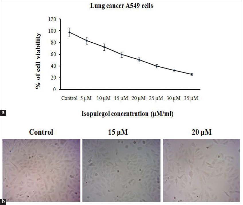
Figure 1a demonstrates that the isopulegol substantially inhibited the A549 cell viability in a dose-dependent manner. In other words, increasing the concentration of isopulegol also resulted in an appropriate increase in inhibitory effects on the growth of A549 cells. Isopulegol (20 μM/mL) was observed to inhibit 50% of A549 cell growth after test in various concentrations (5–35 μM/mL). Therefore, the half maximal inhibitory concentration (IC50) was fixed at 20 μM/mL for further experiments utilized in additional investigations.

thumblarge

Figure :1 Effects of isopulegol on A549 cell viability: (a) Treated with various concentrations of isopulegol and evaluation using MTT assay and (b) morphological observation of cells after isopulegol treatment for 24 h

Effect of Isopulegol on A549 Cell Morphology

The synergistic effect of isopulegol on the morphology of A549 cells was assessed. Isopulegol affects changes in the morphology of A549 cells, as highlighted by changes in the shape of cells. A decrease in the cell number (50%) was also observed. In addition, isopulegol (15–20 μM/mL) treated A549 cells have significant morphological changes, including shape variations, cell shrinkages, and cell detachments [Figure 1b].

Effect of Isopulegol on Intracellular ROS in A549 Cells

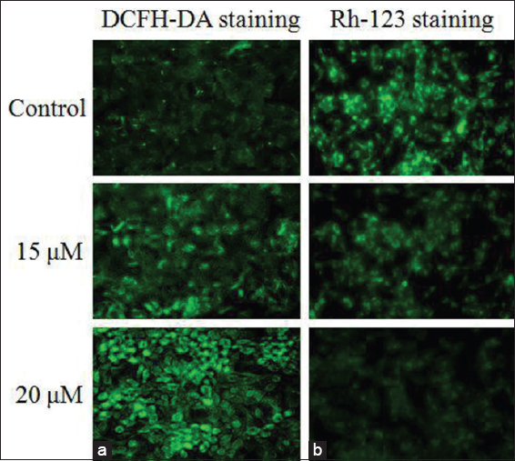
The improvement of intracellular ROS production on the A549 cells was examined using DCFH-DA fluorescence staining. Figure 2a shows that the isopulegol (15 and 20 μM/mL) noticeably increased the intracellular ROS production in the A549 cells. A bright green fluorescence reveals the enhanced accumulation of intracellular ROS, promoting the apoptotic process in the isopulegol treated A549 cells. The untreated control cells exhibit none of the green fluorescence that indicates the excess generation of intracellular ROS in A549 cells.

thumblarge

Figure :2 Effects of isopulegol on intracellular ROS and MMP in A549 cells (treated with isopulegol for 24 h). (a) ROS detection through DCFH-DA staining and (b) MMP evaluation is using Rh-123 staining

Effect of Isopulegol on Mitochondrial Membrane Potential (Δψm) in A549 Cells

The isopulegol stimulated alterations in the Δψm of A549 cells were examined using the Rh-123 fluorescence staining. The increased status of Δψm of A549 cells exhibits bright green fluorescence while the diminished levels of Δψm indicate weak green fluorescence. Figure 2b demonstrates that the control cells have strong green fluorescence; alternatively, the isopulegol treated A549 cells have weak green fluorescence. The later evidentiary exhibits the decreased value of Δψm of A549 cells. Therefore, it is obvious that isopulegol has the capability to reduce the Δψm of lung cancer cells.

Effect of Isopulegol on Apoptotic Induction in A549 Cells

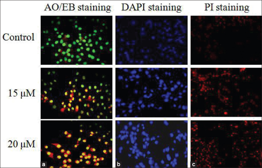
Isopulegol was also assessed to detect the apoptosis stimulating effect on the A549 cancer cells by dual (AO/EB) staining technique. The AO/EB fluorescence staining method was used to find the apoptosis linked nuclear damages during the apoptotic process. Figure 3a represents that the untreated control cells displayed the AO stained green fluorescence. Isopulegol treated A549 cells show an intense EB stained orange fluorescence that indicates the late apoptotic stage cells. Hence, the test proves that isopulegol has the potential to stimulate apoptosis in lung cancer cells. Both concentrations (15 and 20 μM/mL) of the isopulegol have induced apoptosis in the lung cancer A549 cells and it is confirmed by the intense orange fluorescence, compared to the control cells.

thumblarge

Figure :3 (a-c) Effects of isopulegol induced apoptosis in A549 cells treated with various concentrations of isopulegol (15 and 20 μM/ml) for 24 h and the nuclei stained by AO/EB, DAPI, and PI

Effect of Isopulegol on Changes of Nuclear Morphology of A549 Cells

Nuclear fragmentation of the A549 cells was analyzed using DAPI staining assay. Figure 3b shows that the isopulegol has noticeably altered the nuclear morphology as well as the quantity of A549 cells. The bright blue fluorescence has been noted in the A549 cells that are treated with isopulegol compared to the control A549 cells. Moreover, isopulegol treatment efficiently changed the nuclear content, promoting A549 cell death. Therefore, earlier observed nuclear changes indicate that isopulegol can be used inhibits of lung cancer cells.

Effect of Isopulegol-Induced Apoptosis on A549 Cells

The propidium iodide (PI) is a fluorescence staining and widely employed to differentiate the necrotic cells from viable cells. PI cannot penetrate the membrane of viable cells, thereby, only apoptotic or necrotic cells take up the PI staining. Figure 3c shows an intense red fluorescence in the morphology of isopulegol treated A549 cells compared to the untreated control cells. The intensity of the red fluorescence may be indicative of a large number of apoptotic cells. Hence, it is clear that the isopulegol treatment has stimulated necrosis in the A549 cells.

Effect of Isopulegol on SOD, CAT, GSH, and TBARS Levels in A549 Cells

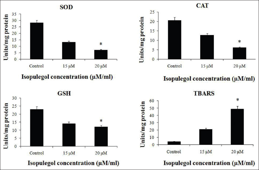
The effects of isopulegol on the action of antioxidant enzymes are shown in Figure 4. The treatment of A549 cells using isopulegol results in a noticeable reduction in antioxidant enzymes (SOD, CAT, and GSH). Besides, isopulegol also encourages the production of LPO product (TBARS) in A549 cells. The results prove that isopulegol can promote free radicals mediated apoptosis in A549 cells.

thumblarge

Figure :4 Effects of isopulegol of antioxidant enzymes action on A549 cells treated with different concentrations (15 and 20 μM/ml) of isopulegol

Effect of Isopulegol on Apoptotic Protein Expression in A549 Cells

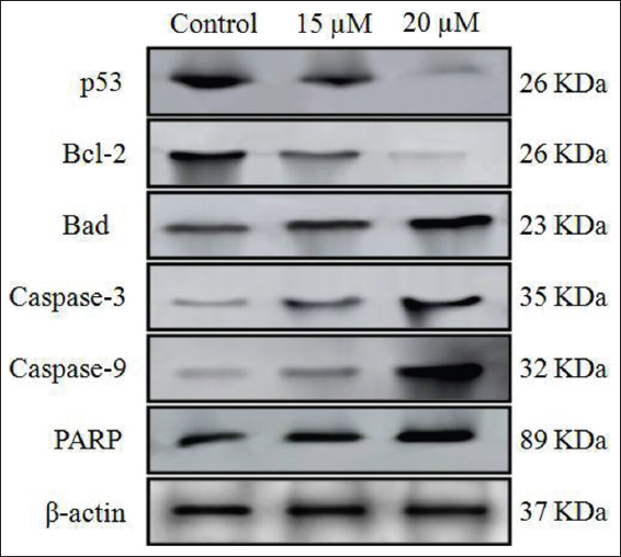
The Western blot study of isopulegol treated A549 cells reveals the status of apoptotic protein expressions, that is, p53, Bad, caspase-3 and-9, and PARP. These proteins are the major proteins, which are responsible for cell apoptosis. Figure 5 shows that isopulegol treatment has significantly enhanced the expression level of apoptotic proteins in the A549 cells. It proves that the isopulegol can stimulate apoptosis in human lung sarcoma cells.

thumblarge

Figure :5 Effects of isopulegol on the expression of apoptotic markers in A549 cells. (a) Treated with various concentrations of isopulegol (15 and 20 μM/mL) and detection of the expressions of p53, Bad, Bcl-2, caspase-3 and caspase-9, PAP, and β-actin by Western blot and (b) the intensity of the bands expressed as optical density (OD) analysis (data presented as mean ± SD and *P < 0.05 relative to control group)

Effect of Isopulegol on the Expression of Inflammatory Modulators in A549 Cells

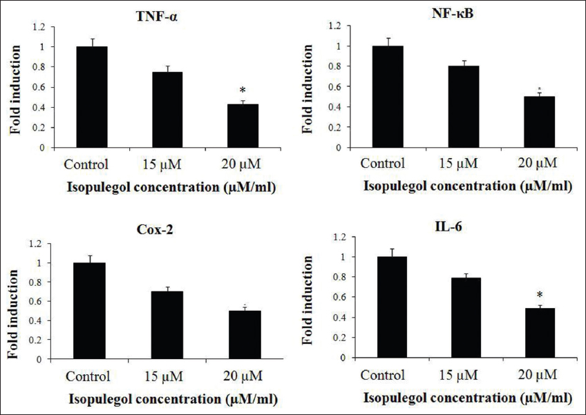
The expression status of mRNA of inflammatory regulators, that is, tumor necrosis factor-alpha (TNF-α), nuclear factor-kappa B (NF-kB), interleukin-6 (Il-6), and cyclooxygenase-2 (Cox-2) in the A549 cells was investigated through the RT-PCR technique. Figure 6 demonstrates that isopulegol treated A549 cells have a noticeable reduction in the expression status of mRNA of inflammatory regulators than the untreated control cells [Figure 6].

thumblarge

Figure :6 Effects of isopulegol on the mRNA expression of inflammatory markers in A549 cells. Isopulegol treated with various concentrations (15 and 20 μM/mL) of TNF-α, NF-kB, IL-6, and Cox-2 detected by RT-PCR

DISCUSSION

Lung cancer is the second most dangerous cancers accountable for numerous cancer-associated deaths worldwide.[33] In general, if the pulmonary sarcoma is diagnosed at a very late stage, many lung cancer incidences are no longer treatable by remedial methods. Cort et al.[34] demonstrated that the multiple redox – various mechanisms implicated in multidrug resistance (MDR) prompted signifying redox-active drugs (antioxidants and pro-oxidants) or suppressor of inducible antioxidant resistance as a novel approach to decreased MDR. Cell death can happen if the cells get exposed to cytotoxic causes.[35] Normally, two kinds of cells are detected frequently, that is, apoptosis and necrosis. The apoptotic cells are affected by the various molecular and cellular mechanisms such as condensation, fragmentation of DNA, blebbing of membranes, and generation of apoptotic bodies. To investigate the apoptotic stimulating agents, a fluorescence microscopic examination of several stained techniques was utilized.[12,36] The determination of the apoptotic level was often carried out through fluorescence microscopy, which reveals the image of apoptotic bodies and condensed chromatin compared to non-apoptotic cells.[36-38]

Oxidative stress condition is a dysfunction of cells that occurs due to the accumulation of excessive ROS in the cells, leading to cell damage and cell death. To reduce cell damages due to excess ROS, free radicals must be converted into other non-toxic substances, it is commonly done using antioxidant enzymes in normal cells. There is a need to suppress the level of the antioxidant enzyme, which eventually leads to elevated ROS accumulation, thereby resulting in apoptosis in cancer cells.[39,40] In the same way, A549 cells treated with isopulegol have markedly suppressed levels of antioxidant enzymes (CAT, SOD, and GSH) and eventually elevated levels of the LPO product (TBARS).

The dual (AO/EB) fluorescence staining technique was widely used to distinguish the nuclear damages due to apoptosis, in which the stained cells were examined with a fluorescence microscope. It can distinctly differentiate the various stages of apoptosis.[41] The DAPI staining is commonly used to detect the DNA fragmentation and nuclear condensation, which is directly linked to the apoptotic process.[42] The level of intracellular ROS synthesis is inspected using the fluorescence dye DCFH-DA and the MMP level of the treated cells can be visualized through the Rh-123 staining method.[43] In the present study, the results of fluorescence staining assays demonstrate that the isopulegol effectively induces apoptosis through elevated ROS accretion and reduced MMP in the A549 cells.

Interestingly, Dragovich et al.[44] highlighted that the treatment with imexon, to moderate metastatic cancer and also reduction on the GSH status in the cells, thereby promoted the overaccumulation of ROS in the patients. Besides, the treatment with mangafodipir, a novel adjuvant chemotherapeutic agent, particularly blocked the SOD in cancer cells, thus, promoting the discharge of H2O2 radicals in cells.[45] Similarly, isopulegol possesses the anticonvulsant effect on the pentylenetetrazol-induced mouse model which increases the activity of antioxidants (CAT and GSH) and LPO in brain hippocampus of mice. Moreover, chemotherapeutic agents target excess accumulation of ROS in cancer cells that were proved on both experimental and pre-clinical investigations.[46]

The cells under oxidative stress condition promote the expression level of p53 genes that eventually secrete ROS for cell apoptosis. Conversely, in the normal cells, it reduces the secretion of ROS, thereby, guarding the cells against severe effects of ROS. Due to this dual action, p53 was regarded as a double-edged sword, in additional, it is known as a tumor-suppressing gene for its effects on inhibiting the ROS and promoting the transforming growth factor.[47,48] Reducing the uninhibited cell growth of tumors has been regarded as a possible way of healing cancer by utilizing the natural cell necrotic mechanism. Furthermore, promoting apoptosis in malignant cells constitutes a promising non-surgical or non-radiotherapy approach in cancer treatment. The application of apoptosis in cancer treatment has the potential of treating all kinds of sarcomas. It was already highlighted that numerous new anti-cancer agents targeted the several pathways of both intrinsic and extrinsic mechanisms of apoptosis in sarcoma cells.[49,50] Dias et al.[21] reported that (-) isopulegol epoxide coated bioinspired-metalloporphyrins magnetic nanocomposites suppress the growth of human osteosarcoma (MG-63) cells.

The stimulation of caspase proteins and BAD proteins is an essential approach in the apoptotic mechanism that is mediated through Bcl-2 types. The caspases are identified as dormant state zymogen and undergo proteolytic stimulation during the apoptotic process. Caspase-9 is a mediator in the caspase and contributes to the mitochondrial regulated inherent apoptotic pathway; also, caspase-3 is a prime factor that prompts the investigation of apoptosis.[51] As depicted in Figure 6, the expression status of caspase-3 and caspase-9 has significantly elevated the expression status of isopulegol treated A549 cells. Besides, it suppressed the anti-apoptotic protein, that is, Bcl-2 expression level in A549 cells. Hence, it proves that isopulegol can stimulate apoptosis in A549 cells. The PARP has an essential role in the protection of DNA as well as a substrate for the caspase-3 that contributes to apoptosis. PARP gets broken-down through caspase-3 that directs DNA fragments in apoptosis. We noted that isopulegol treatment results in cleaved subsets in A549 cells. Thus, the results of the present research have proven that the isopulegol can commence and promote apoptosis in A549 cells. Besides, a marked reduction in the survival of A549 cells has been noticed after isopulegol treatment and it may be due to the apoptosis induction capacity of isopulegol. The DAPI staining cancer cells and Western blotting examination of apoptotic protein expression of isopulegol treated A549 cells have confirmed the same result.

The NF-kB signaling is a highly imperative pathway for the stimulation of transcription of genes for encoding the proteins that contribute to complicated apoptotic mechanisms in cells.

The triggered NF-kB has promoted the transcription range of anti-apoptotic proteins, for example, Bcl-2 which results in the resistance to the chemotherapeutic apoptotic system.[52,53] Thus, it is necessary to suppress the NF-kB status in the cancer cells. Moreover, the result of RT-PCR analysis confirmed that the isopulegol treatment has noticeably suppressed the mRNA expression of NF-kB and other anti-inflammatory markers, for example, TNF-α, IL-6, and Cox-2 in the human lung adenocarcinoma (A549) cells, which proved the therapeutic values of isopulegol.

CONCLUSIONS

The potential benefit of isopulegol as a therapeutic agent that targets cell proliferative mechanisms in A549 cells has been demonstrated in the present study. The findings of the present research study revealed that the isopulegol was highly effective against the A549 cells. Isopulegol treatment diminished the antioxidant shielding system and elevated the intercellular ROS accumulation, therefore, promoting apoptotic cell death in lung cancer cells. Isopulegol also increases levels of TNF-α and NF-kB in cells. The levels of earlier inflammatory mediators were significantly modulated in the induced A549 cancer cells, which could be related to the ROS increased on the induction of apoptosis. Accordingly, it is concluded that isopulegol can be a promising chemotherapeutic agent toward the treatment of lung cancer.

REFERENCES

1.  Kumar R, Lu SK, Minchom A, Sharp A, Davidson M, Gunapala R, et al. A phase 1b trial of the combination of an all-oral regimen of capecitabine and erlotinib in advanced non-small cell lung cancer in Caucasian patients. Cancer Chemother Pharmacol 2016;77:375-83.

2.  Falzone L, Salomone S, Libra M. Evolution of cancer pharmacological treatments at the turn of the third millennium. Front Pharmacol 2018;9:1300.

3.  Blum HE. Hepatocellular carcinoma:Therapy and prevention. World J Gastroenterol 2005;11:7391-400.

4.  Luo D, Carter KA, Miranda D, Lovell JF. Chemophototherapy:An emerging treatment option for solid tumors. Adv Sci (Weinh) 2016;4:1600106.

5.  Jin J, Chen J, Suo S, Qian W, Meng H, Mai W, et al. Low-dose cytarabine, aclarubicin and granulocyte colony-stimulating factor priming regimen versus idarubicin plus cytarabine regimen as induction therapy for older patients with acute myeloid leukemia. Leuk Lymphoma 2015;56:1691-7.

6.  Szwed M, Laroche-Clary A, Robert J, Jozwiak Z. Efficacy of doxorubicin-transferrin conjugate in apoptosis induction in human leukemia cells through reactive oxygen species generation. Cell Oncol (Dordr) 2016;39:107-18.

7.  Chow R, Tsao M, Chiu L, Popovic M, Milakovic M, Lam H, et al. Efficacy of the combination neurokinin-1 receptor antagonist, palonosetron, and dexamethasone compared to others for the prophylaxis of chemotherapy-induced nausea and vomiting:A systematic review and meta-analysis of randomized controlled trials. Ann Palliat Med 2018;7:221-33.

8.  Hail N Jr., Carter BZ, Konopleva M, Andreeff M. Apoptosis effector mechanisms:A requiem performed in different keys. Apoptosis 2006;11:889-904.

9.  Abraham MC, Shaham S. Death without caspases, caspases without death. Trends Cell Biol 2004;14:184-93.

10.  Halliwell B. Oxidative stress in cell culture:An under-appreciated problem?FEBS Lett 2003;5403-6.

11.  Ozben T. Oxidative stress and apoptosis:Impact on cancer therapy. J Pharm Sci 2007;96:2181-96.

12.  Dey D, Das S, Yadav R, Ranjani A, Gyathri L, Roy SC, et al. Design of a mononuclear copper (II)-phenanthroline complex:Catechol oxidation, DNA cleavage and antitumor properties. Polyhedron 2016;106:106-14.

13.  Lee KH. Discovery and development of natural product-derived chemotherapeutic agents based on a medicinal chemistry approach. J Nat Prod 2010;73:500-16.

14.  Remila S, Atmani-Kilani D, Delemasure S, Connat J, Azib L. Antioxidant cytoprotective, anti-inflammatory and anticancer activities of Pistacia lentiscus (Anacardiaceae) leaf and fruit extracts. Eur J Integr Med 2015;7:274-86.

15.  Newman DJ, Cragg GM, Snader KM. Natural products as sources of new drugs over the period 1981-2002. J Nat Prod 2003;66:1022-37.

16.  Paik SY, Koh KH, Beak SM, Paek SH, Kim JA. The essential oils from Zanthoxylum schinifolium pericarp induce apoptosis of HepG2 human hepatoma cells through increased production of reactive oxygen species. Biol Pharm Bull 2005;28:802-7.

17.  Vernin GA, Parkanyi C, Cozzolino F, Fellous R. GC/MS Analysis of the Volatile Constituents of Corymbia citriodora Hook. From Réunion Island. J Essent Oil Res 2004;16:560-5.

18.  Bounihi A, Hajjaj G, Alnamer R, Cherrah Y, Zellou A. In vivo potential anti-inflammatory activity of Melissa officinalis L. essential oil. Adv Pharmacol Sci 2013;2013:101759.

19.  Rao BR, Kaul PN, Syamasundar KV, Ramesh S. Comparative composition of decanted and recovered essential oils of Eucalyptus citriodora Hook. Flavour Fragr J 2003;18:133-5.

20.  Silva MI, Silva MA, de Aquino Neto MR, Moura BA, de Sousa HL, de Lavor EP, et al. Effects of isopulegol on pentylenetetrazol-induced convulsions in mice:Possible involvement of GABAergic system and antioxidant activity. Fitoterapia 2009;80:506-13.

21.  Dias LD, de Carvalho AL, Pinto SM, Aquino GL, Calvete MJ, Rossi LM, et al. Bioinspired-metalloporphyrin magnetic nanocomposite as a reusable catalyst for synthesis of diastereomeric (-)-isopulegol epoxide:Anticancer activity against human osteosarcoma cells (MG-63). Molecules 2018;24:52.

22.  Silva MI, Moura BA, Neto MR, ToméAda R, Rocha NF, de Carvalho AM, et al. Gastroprotective activity of isopulegol on experimentally induced gastric lesions in mice:Investigation of possible mechanisms of action. Naunyn Schmiedebergs Arch Pharmacol 2009;380:233-45.

23.  Próspero DF, Filho AC, Piauilino CA, Lopes EM, de Sousa DP, de Castro Almeida FR. Effects of isopulegol in acute nociception in mice:Possible involvement of muscarinic receptors, opioid system and l-arginine/NO/cGMP pathway. Chem Biol Interact 2018;293:55-60.

24.  Oliveira Fde A, Andrade LN, de Sousa EB, de Sousa DP. Anti-ulcer activity of essential oil constituents. Molecules 2014;19:5717-47.

25.  Ames BN, Mccann J, Yamasaki E. Methods for detecting carcinogens and mutagens with the Salmonella/mammalian-microsome mutagenicity test. Mutat Res 1975;31:347-64.

26.  Mosmann T. Rapid colorimetric assay for cellular growth and survival:Application to proliferation and cytotoxicity assays. J Immunol Methods 1983;65:55-63.

27.  Merlin NJ, Parthasarathy V, Santhoshkumar TR. Induction of apoptosis in huma breast cancer cell line MCF-7 by phytochemicals from Gmelina asiatica. Afr J Biotech 2010;9:4451-6.

28.  Eskandani M, Hamishehkar H, Dolatabadi JE. Cytotoxicity and DNA damage properties of tert-butylhydroquinone (TBHQ) food additive. Food Chem 2004;153:315-20.

29.  Sun YI, Oberley LW, Li Y. A simple method for clinical assay of superoxide dismutase. Clin Chem 1988;34:497-500.

30.  Góth L. A simple method for determination of serum catalase activity and revision of reference range. Clin Chim Acta 1991;196:143-51.

31.  Rotruck JT, Pope AL, Ganther HE, Swanson AB, Hafeman DG, Hoekstra WG. Selenium:Biochemical role as a component of glutathione peroxidase. Science 1973;179:588-90.

32.  Buege JA, Aust SD. Microsomal lipid peroxidation. Methods Enzymol 1978;52:302-10.

33.  Maucort-Boulch D, de Martel C, Franceschi S, Plummer M. Fraction and incidence of liver cancer attributable to hepatitis B and C viruses worldwide. Int J Cancer 2018;142:2471-7.

34.  Cort A, Ozben T, Saso L, De Luca C, Korkina L. Redox control of multidrug resistance and its possible modulation by antioxidants. Oxid Med Cell Longev 2016;2016:4251912.

35.  Shigematsu H, Yoshida K, Sanada Y, Osada S, Takahashi T, Wada Y, et al. Rapamycin enhances chemotherapy-induced cytotoxicity by inhibiting the expressions of TS and ERK in gastric cancer cells. Int J Cancer 2010;126:2716-25.

36.  Saha SK, Sikdar S, Mukherjee A, Bhadra K, Boujedaini N, Khuda-Bukhsh AR. Ethanolic extract of the goldenseal, Hydrastis canadensis, has demonstrable chemopreventive effects on HeLa cells in vitro:Drug-DNA interaction with calf thymus DNA as target. Environ Toxicol Pharmacol 2013;36:202-14.

37.  Ansil PN, Wills PJ, Varun R, Latha MS. Cytotoxic and apoptotic activities of Amorphophallus campanulatus (Roxb.) Bl. tuber extracts against human colon carcinoma cell line HCT-15. Saudi J Biol Sci 2014;21:524-31.

38.  Peng CH, Huang CN, Hsu SP, Wang CJ. Penta-acetyl geniposide induce apoptosis in C6 glioma cells by modulating the activation of neutral sphingomyelinase-induced p75 nerve Growth factor receptor and protein kinase Cdelta pathway. Mol Pharmacol 2006;70:997-1004.

39.  Niska K, Santos-Martinez MJ, Radomski MW, Inkielewicz-Stepniak I. CuO nanoparticles induce apoptosis by impairing the antioxidant defense and detoxification systems in the mouse hippocampal HT22 cell line:Protective effect of crocetin. Toxicol In Vitro 2015;29:663-71.

40.  He L, He T, Farrar S, Ji L, Liu T, Ma X. Antioxidants maintain cellular redox homeostasis by elimination of reactive oxygen species. Cell Physiol Biochem 2015;44:532-53.

41.  Gherghi IC, Girousi ST, Voulgaropoulos AN, Tzimou-Tsitouridou R. Study of interactions between DNA-ethidium bromide (EB) and DNA-acridine orange (AO), in solution, using hanging mercury drop electrode (HMDE). Talanta 2003;61:103-12.

42.  Vandghanooni S, Forouharmehr A, Eskandani M, Barzegari A, Kafil V, Kashanian S, et al. Cytotoxicity and DNA fragmentation properties of butylated hydroxyanisole. DNA Cell Biol 2013;32:98-103.

43.  Zhou GZ, Shi YY, Cui LS, Li AF, Wang QQ, Liu M. Oxymatrine induces A549 human nonsmall lung cancer cell apoptosis via extrinsic and intrinsic pathways. Mol Med Rep 2018;17:1071-6.

44.  Dragovich T, Gordon M, Mendelson D, Wong L, Modiano M, Chow HH, et al. Phase I trial of imexon in patients with advanced malignancy. J Clin Oncol 2007;25:1779-84.

45.  Alexandre J, Nicco C, Chéreau C, Laurent A, Weill B, Goldwasser F, et al. Improvement of the therapeutic index of anticancer drugs by the superoxide dismutase mimic mangafodipir. J Natl Cancer Inst 2006;98:236-44.

46.  Xiao D, Powolny AA, Moura MB, Kelley EE, Bommareddy A, Kim SH, et al. Phenethyl isothiocyanate inhibits oxidative phosphorylation to trigger reactive oxygen species-mediated death of human prostate cancer cells. J Biol Chem 2010;285:26558-69.

47.  Das L, Vinayak M. Long term effect of curcumin in restoration of tumour suppressor p53 and phase-II antioxidant enzymes via activation of Nrf2 signalling and modulation of inflammation in prevention of cancer. PLoS One 2015;10:e0124000.

48.  Gong Z, Yang Q, Zeng Z, Zhang W, Li X, Zu X, et al. An integrative transcriptomic analysis reveals p53 regulated miRNA, mRNA, and lncRNA networks in nasopharyngeal carcinoma. Tumour Biol 2013;37:3683-95.

49.  Villa-Pulgarín JA, Gajate C, Botet J, Jimenez A, Justies N, Varela M, et al. Mitochondria and lipid raft-located FOF1-ATP synthase as major therapeutic targets in the antileishmanial and anticancer activities of ether lipid edelfosine. PLoS Negl Trop Dis 2017;11:e0005805.

50.  Bao H, Zhang Q, Zhu Z, Xu H, Ding F, Wang M, et al. BHX, a novel pyrazoline derivative, inhibits breast cancer cell invasion by reversing the epithelial-mesenchymal transition and down-regulating Wnt/b-catenin signalling. Sci Rep 2017;7:9153.

51.  Shang HS, Chang CH, Chou YR, Yeh MY, Au MK, Lu HF, et al. Curcumin causes DNA damage and affects associated protein expression in HeLa human cervical cancer cells. Oncol Rep 2016;36:2207-15.

52.  Yang HJ, Wang M, Wang L, Cheng BF, Lin XY, Feng ZW. NF-kB regulates caspase-4 expression and sensitizes neuroblastoma cells to Fas-induced apoptosis. PLoS One 2015;10:e0117953.

53.  Zheng HC. The molecular mechanisms of chemoresistance in cancers. Oncotarget 2017;8:59950-64.